|

by Joel M. Moskowitz, Ph.D.
BERKELEY, Calif.
September 23, 2013
from
StopSmartMetres Website
New peer-reviewed
research finds that
30 minutesí exposure to LTE
[4G] cellphone radiation
affects brain activity on both
sides of the brain.
PRLog - Press Release
The first study on the short-term
effects of Long Term Evolution (LTE), the fourth generation
cell phone technology, has been published online in the
peer-reviewed journal, Clinical Neurophysiology. (1)

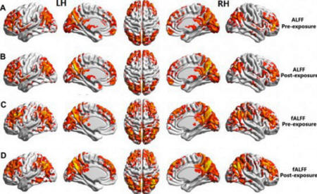
Brain images
pre- and post-LTE
(4G) exposure
In a controlled experiment, researchers exposed the right ear of 18
participants to LTE cell-phone radiation for 30 minutes.
The source of the radiation was 1
centimeter from the ear, and the absorbed amount of radiation in the
brain was well within international (ICNIRP) cell phone legal
limits. The researchers employed a double-blind, crossover,
randomized and counter-balanced design to eliminate any possible
study biases.
The resting state brain activity of each
participant was measured by magnetic resonance imaging (fMRI)
at two times - after exposure to LTE microwave radiation, and after
a sham exposure.
The results demonstrated that LTE exposure affected brain neural
activity not only in the closer brain region but also in the remote
region, including the left hemisphere of the brain. The study helps
explain the underlying neural mechanism for the remote effects of
microwave radiation in the brain.
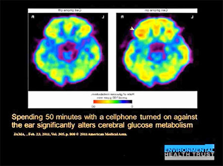
In 2011, Dr. Nora Volkow, Director of the National Institute
on Drug Abuse, published a similar study in the Journal of the
American Medical Association that received worldwide news coverage.
Dr. Volkow reported that a 50 minute
exposure to CDMA, a second generation cell phone technology,
increased brain activity in the region of the brain closest to the
cell phone. (2)
The current study establishes that short-term exposure to LTE
microwave radiation affects the usersí brain activity.
Although LTE is too new for the
long-term health consequences to have been studied, we have
considerable evidence that long-term cell phone use is associated
with various health risks including increased risk of head and neck
cancers, sperm damage, and reproductive health consequences for
offspring (i.e., ADHD).
Cell phone users, especially pregnant women and children, should
limit their cell phone use. Moreover, cell phone users should not
keep their phones near their head, breasts or reproductive organs
when using the phone or whenever the phone is turned on unless it is
in airplane mode.
For more information about the health effects of cell phone
radiation see
Electromagnetic Radiation Safety Web site.
References
(1) - Bin Lv, Zhiye Chen, Tongning
Wu, Qing Shao, Duo Yan, Lin Ma, Ke Lu, Yi Xie. The alteration of
spontaneous low frequency oscillations caused by acute
electromagnetic fields exposure. Clinical Neurophysiology.
Published online 4 September 2013.
http://www.ncbi.nlm.nih.gov/pubmed/24012322
Abstract
Objective The motivation of this study is to evaluate the
possible alteration of regional resting state brain activity
induced by the acute radiofrequency electromagnetic field (RF-EMF)
exposure (30 min) of Long Term Evolution (LTE) signal.
Methods We designed a controllable near-field LTE RF-EMF
exposure environment. Eighteen subjects participated in a
double-blind, crossover, randomized and counterbalanced
experiment including two sessions (real and sham exposure). The
radiation source was close to the right ear.
Then the resting state fMRI signals
of human brain were collected before and after the exposure in
both sessions. We measured the amplitude of low frequency
fluctuation (ALFF) and fractional ALFF (fALFF) to characterize
the spontaneous brain activity.
Results We found the decreased ALFF value around in left
superior temporal gyrus, left middle temporal gyrus, right
superior temporal gyrus, right medial frontal gyrus and right
paracentral lobule after the real exposure. And the decreased
fALFF value was also detected in right medial frontal gyrus and
right paracentral lobule.
Conclusions The study provided the evidences that 30 min LTE
RF-EMF exposure modulated the spontaneous low frequency
fluctuations in some brain regions.
Significance With resting state fMRI, we found the alteration of
spontaneous low frequency fluctuations induced by the acute LTE
RF-EMF exposure.
(2) - Volkow ND, Tomasi D, Wang GJ,
Vaska P, Fowler JS, Telang F, Alexoff D, Logan J, Wong C.
Effects of cell phone radiofrequency signal exposure on brain
glucose metabolism. JAMA. 2011 Feb 23;305(8):808-13. doi:
10.1001/jama.2011.186.
http://www.ncbi.nlm.nih.gov/pmc/articles/PMC3184892/
Abstract
CONTEXT
The dramatic increase in use of
cellular telephones has generated concern about possible
negative effects of radiofrequency signals delivered to the
brain.
However, whether acute cell phone
exposure affects the human brain is unclear.
OBJECTIVE
To evaluate if acute cell phone
exposure affects brain glucose metabolism, a marker of brain
activity.
DESIGN, SETTING, AND PARTICIPANTS
Randomized crossover study conducted
between January 1 and December 31, 2009, at a single US
laboratory among 47 healthy participants recruited from the
community.
Cell phones were placed on the left
and right ears and positron emission tomography with ((18)F)
fluorodeoxyglucose injection was used to measure brain glucose
metabolism twice, once with the right cell phone activated
(sound muted) for 50 minutes ("on" condition) and once with both
cell phones deactivated ("off" condition).

Statistical parametric mapping was
used to compare metabolism between on and off conditions using
paired t tests, and Pearson linear correlations were used to
verify the association of metabolism and estimated amplitude of
radiofrequency-modulated electromagnetic waves emitted by the
cell phone.
Clusters with at least 1000 voxels
(volume >8 cm(3)) and P < .05 (corrected for multiple
comparisons) were considered significant.
MAIN OUTCOME MEASURE:
Brain glucose metabolism computed as
absolute metabolism (μmol/100 g per minute) and as normalized
metabolism (region/whole brain).
RESULTS:
Whole-brain metabolism did not
differ between on and off conditions. In contrast, metabolism in
the region closest to the antenna (orbitofrontal cortex and
temporal pole) was significantly higher for on than off
conditions (35.7 vs. 33.3 μmol/100 g per minute; mean
difference, 2.4 [95% confidence interval, 0.67-4.2]; P = .004).
The increases were significantly
correlated with the estimated electromagnetic field amplitudes
both for absolute metabolism (R = 0.95, P < .001) and normalized
metabolism (R = 0.89; P < .001).
CONCLUSIONS:
In healthy participants and compared
with no exposure, 50-minute cell phone exposure was associated
with increased brain glucose metabolism in the region closest to
the antenna.
This finding is of unknown clinical
significance.
|


