|

by
Dr. Denis G. Rancourt, PhD
June 02, 2020
from
ResearchGate Website
PDF format

Summary / Abstract
The latest data of all-cause mortality by week does not show a
winter-burden mortality that is statistically larger than for past
winters.
There was no plague...
However, a sharp "COVID peak" is
present in the data, for several jurisdictions in Europe and the
USA.
This all-cause-mortality "COVID peak" has unique
characteristics:
-
Its sharpness, with a full-width at half-maximum of only
approximately 4 weeks;
-
Its lateness in the infectious-season cycle, surging after week-11
of 2020, which is unprecedented for any large sharp-peak feature;
-
The synchronicity of the onset of its surge, across continents, and
immediately following the WHO declaration of the pandemic;
-
and its USA state-to-state absence or presence for the same viral
ecology on the same territory, being correlated with nursing home
events and government actions rather than any known viral strain
discernment.
These "COVID peak" characteristics, and a review of the
epidemiological history, and of relevant knowledge about viral
respiratory diseases, lead me to postulate that the "COVID peak"
results from,
an accelerated mass homicide of immune-vulnerable
individuals, and individuals made more immune-vulnerable, by
government and institutional actions, rather than being an
epidemiological signature of a novel virus, irrespective of the
degree to which the virus is novel from the perspective of viral
speciation.
The paper is organized into the following sections:
-
Cause-of-death-attribution data is intrinsically unreliable
-
Year-to-year winter-burden mortality in mid-latitude nations is
robustly regular
-
Why is the winter-burden pattern of mortality so regular and
persistent?
-
A simple model of viral respiratory disease de facto virulence
-
All-cause mortality analysis of
COVID-19
-
Interpreting the all-cause mortality "COVID peak"
Cause-of-death-attribution data is intrinsically unreliable
Assignment of cause of death, with infectious diseases and
comorbidity, is not only technically difficult (e.g., Simonsen et
al., 1997; Marti-Soler et al., 2014) but also contaminated by
physician-bias, politics and news media.
This has been known since modern epidemiology was first practiced.
Here is Langmuir (1976) quoting the renowned pioneer
William Farr,
regarding the influenza epidemic of 1847:
Farr uses this epidemic to chide physicians mildly on their narrow
views pointing out that sharp increases were observed not only in
influenza itself but in bronchitis, pneumonia and asthma and many
other non-respiratory causes, he states:
'... there is a strong disposition among some English practitioners
not only to localize disease but to see nothing but the local
disease.
Hence, although it is certain that the high mortality on
record was the immediate result of the epidemic of influenza, the
deaths referred to that cause are only 1,157.'
And, such bias is generally recognized by leading epidemiologists (Lui
and Kendal, 1987):
... the decision to classify deaths into "pneumonia and influenza"
is subjective and potentially inconsistent.
On one hand, the effect
of influenza or influenza-related pneumonia may be underestimated
because underlying chronic diseases, particularly in the elderly,
are usually noted as the cause of death on the death certificate.
On
the other hand, after influenza activity has been publicly reported
there may be an increased tendency to classify deaths as due to
"pneumonia and influenza," thereby amplifying the rate of increase
in P&I deaths or, when a decline in influenza activity is reported,
a bias toward decreasing the classification of deaths related to
"pneumonia and influenza" may result.
Surveys to evaluate these
possibilities have not been done.
One can reasonably expect that in the current world of social media,
with a World-Health-Organization-declared (WHO-declared) "pandemic",
such bias will only be greater compared to its presence in past
viral respiratory disease epidemics.
For example, it is difficult to interpret the synchronicity of
the
WHO declaration of COVID-19 as a pandemic and the onset of the
observed surge in reported COVID-19 cases and deaths as being the
product of either coincidence or extraordinary forecasting ability
of the global health-monitoring system:

Figure 1:
Globally reported COVID-19 cases,
and reported
COVID-19-assigned deaths, by day.
WHO data was accessed on 30 May 2020.
The vertical lines in pencil
indicate the date at which
the WHO declared the pandemic.

Figure 2:
Globally reported new COVID-19 cases per day,
by
continent. WHO datawas accessed on 30 May 2020.
The vertical line in pencil indicates
the date at which the WHO
declared the pandemic.
Instead, in light of past epidemics, it is more likely that this
remarkable synchronicity phenomenon arises from biased reporting, in
the flexible context of using urgently manufactured laboratory tests
that are not validated, clinical assessments of a generic array of
symptoms, and tentative cause-of-death assignations of complex
comorbidity circumstances.
That is why rigorous epidemiological studies rely instead on
all-cause mortality data, which cannot be altered by observational
or reporting bias (as discussed in Simonsen et al., 1997; and see
Marti-Soler et al., 2014).
A death is a death is a death...
Year-to-year winter-burden mortality in mid-latitude nations is
robustly regular
Modern human mortality in mid-latitude temperate-climate regions is
robustly seasonal.
Graphs of number of all-cause deaths per unit of
time (month, week, day), in given regions, have a yearly pattern,
with a peak-to-trough amplitude of typically 10% to 30% of the
trough-baseline value, largely irrespective of the specific
pathogens that populate the specific seasons.
High mortality
occurs
in winter, and is thus inverted in the Northern and Southern
hemispheres (e.g., Marti-Soler et al., 2014).
For the USA, the phenomenon is well illustrated in this figure from
Simonsen et al. (1997):

Figure 3:
All-cause mortality, by week,
for the USA, 1972 to 1993
(Simonsen
et al., 1997; from
their Fig. 1).
In such a graph, the area under a peak, to its trough-level
baseline, is the total number of yearly winter-burden deaths above
the trough baseline.
The thus calculated yearly "excess" number of
deaths, here (in the era 1972-1993), is always approximately 8% to
11% of the total yearly trough-baseline-level deaths, also
approximately 8% to 11% of the yearly all-cause mortality.
This regular and seasonal "excess" mortality, or winter burden, has
been an epidemiological challenge to understand, although, starting
with Farr, many epidemiologists originally attributed it almost
entirely to the seasonal influenza-like viral respiratory diseases.
Nonetheless, the agonizing difficulty of understanding the cause(s)
of this remarkably regular and global (both hemispheres, but
inverted) pattern persists, as illustrated by Marti-Soler et al.
(2014) (references omitted):
Given that mortality from cancer showed virtually no seasonality
pattern, the seasonality of overall mortality is driven mostly by
seasonality of both CVD [cardiovascular diseases] and non-CVD/non-cancer
mortality.
For these conditions, and particularly for CVD, exposure
to cold is a plausible explanation for the observed seasonality,
given relationship of cold climate with latitude. Several
longitudinal studies have demonstrated that a decrease in outdoor
temperature was associated with a rise in all-cause mortality.
However, other latitude-dependent factors, such as dietary habits,
sun exposure (vitamin D levels) and human parasitic and infectious
agents might also play a role.
The magnitude of the seasonal pattern for CVD mortality was higher
than that for all-cause mortality. The seasonality of CVD mortality
might be partly due to the joint seasonality of several known CVD
risk factors, as described previously.
Similarly, lifestyle factors
such as diet and physical activity also tend to differ during summer
and winter months.
Moreover, exposure to cold increases energy
expenditure, peripheral vasoconstriction and cardiac afterload, thus
potentially triggering myocardial ischemia 6 and stroke.
Finally,
winter prone influenza infection might also be a trigger for CVD
deaths by exacerbating CVD conditions or due to secondary
complications. This is likely to be the case of concentration of air
pollutants.
The seasonality of non-CVD/non-cancer mortality can relate to the
facts that chronic obstructive pulmonary disease and pneumonia are
frequent diseases in this category and that these disease are
exacerbated by influenza, other influenza-like infections and
concentrations of air pollutants, which are all more frequent in
winter.
A few other diseases in the non-CVD/non-cancer category also
present a seasonal pattern, e.g. depression, suicide, and
oesophageal variceal bleeding.
Why is the winter-burden pattern of mortality so regular and
persistent?
Even the seasonality of the pneumonia and influenza ("P&I") part
alone (which is a large part of what Marti-Soler et al. quantify as
"non-CVD/non-cancer mortality") was not understood until a decade
ago.
Until recently, it was debated whether the P&I yearly pattern
arose primarily because of seasonal change in virulence of the
pathogens, or because of seasonal change in susceptibility of the
host (such as from dry air causing tissue irritation, or diminished
daylight causing vitamin deficiency or hormonal stress).
For example, see Dowell
(2001).
In a sense, the answer is "neither".
In a landmark study, Shaman et al. (2010) showed that the seasonal
pattern of respiratory-disease (P&I) excess mortality can be
explained quantitatively on the sole basis of absolute humidity, and
its direct controlling impact on transmission of airborne pathogens.
Lowen et al. (2007) demonstrated the phenomenon of
humidity-dependent airborne-virus contagiousness in actual disease
transmission between guinea pigs, and discussed potential underlying
mechanisms for the measured controlling effect of humidity.
The underlying mechanism is that the pathogen-laden aerosol
particles or aerosol-size droplets are neutralized within a
half-life that monotonically and significantly decreases with
increasing ambient absolute humidity.
This is based on the seminal
work of Harper (1961).
Harper experimentally showed that
viral-pathogen-carrying droplets were inactivated within shorter and
shorter times, as ambient absolute humidity was increased.
Harper argued that the viruses themselves were made inoperative by
the humidity ("viable decay"), however he admitted that the effect
could be from humidity-enhanced physical removal or gravitational
sedimentation of the droplets ("physical loss"):
"Aerosol viabilities reported in this paper are based on the ratio
of virus titre to radioactive count in suspension and cloud samples,
and can be criticized on the ground that test and tracer materials
were not physically identical."
The latter ("physical loss") seems more plausible to me, since
absolute humidity would have a universal physical effect of causing
particle/droplet growth-by-condensation and gravitational
sedimentation (and, conversely, loss-by-evaporation and
aerosolization), and all tested viral pathogens have essentially the
same humidity-driven "decay".
Furthermore, it is difficult to
understand how a virion (of any virus type) in a droplet would be
molecularly or structurally attacked or damaged by an increase in
ambient humidity.
A "virion" is the complete, infective form of a
virus outside a host cell, with a core of RNA or DNA and a capsid.
No actual molecular or other mechanism of the humidity-driven
intra-droplet "viable decay" of a virion postulated by Harper (1961)
has, to date, been explained or studied, whereas gravitational
sedimentation ("physical loss") is well understood.
In any case, the explanation and model of Shaman et al. (2010) is
not dependent on the particular mechanism of the
absolute-humidity-driven decay of virions in aerosol/droplets.
Shaman's quantitatively demonstrated model of seasonal regional
viral epidemiology is valid for either mechanism (or combination of
mechanisms), whether "viable decay" or "physical loss".
The breakthrough achieved by Shaman et al. is not merely some
academic point. Rather, it has profound health-policy implications,
which have been entirely ignored or overlooked in the current
coronavirus pandemic:
If my view of the mechanism is correct (i.e., "physical loss" rather
than "viable decay"), then:
-
It additionally implies that the transmission vector must be small
aerosol particles in fluid suspension in air, breathed deeply into
the lungs, indoors; not hypothesized routes such as actual fluid or
fomite contact, and not large droplets and spit (that are quickly
gravitationally removed from the air, or captured in the mouth and
digestive system).
-
And it means that social distancing, masks, and handwashing can have
little effect in the actual epidemic spread during the winter season
(see: Rancourt, 2020).
On the epidemiology
modeling side, Shaman's work implies that,
rather than being a fixed number (dependent solely on the
spatial-temporal structure of social interactions in a completely
and variably susceptible population, and on the viral strain), the
epidemic's basic reproduction number (R0) is predominantly dependent
on ambient absolute humidity.
For a definition of R0, see HealthKnowlege-UK (2020):
R0 is "the average number of secondary
infections produced by a typical case of an infection in a
population where everyone is susceptible."
Shaman et al. showed that R0 must be understood to vary seasonally
between humid-summer values of just larger than "1" and dry-winter
values typically as large as "4" (for example, see their Table 2).
In other words, the seasonal infectious viral respiratory diseases
that plague temperate-climate regions every year go from being
intrinsically mildly contagious to virulently contagious, due simply
to the bio-physical mode of transmission controlled by atmospheric
absolute humidity, largely irrespective of any other consideration.
Furthermore, indoor airborne virus concentrations have been shown to
exist (in day-care facilities, health centers, and onboard
airplanes) primarily as aerosol particles of diameters smaller than
2.5 μm, such as in the work of Yang et al. (2011):
"Half of the 16 samples were positive, and their total virus
concentrations ranged from 5,800 to 37,000 genome copies m-3.
On
average, 64 percent of the viral genome copies were associated with
fine particles smaller than 2.5 µm, which can remain suspended for
hours. Modelling of virus concentrations indoors suggested a source
strength of 1.6 ± 1.2 × 10⁵ genome copies m-3 air h-1 and a
deposition flux onto surfaces of 13 ± 7 genome copies m-2 h-1 by
Brownian motion.
Over 1 hour, the inhalation dose was estimated to
be 30 ± 18 median tissue culture infectious dose (TCID50), adequate
to induce infection.
These results provide quantitative support for
the idea that the aerosol route could be an important mode of
influenza transmission."
Such small particles (smaller than 2.5 μm) are part of air fluidity,
are not subject to gravitational sedimentation, and can therefore be
breathed deeply into the lungs.
The next question is:
How many such pathogen-laden particles are
needed to cause infection in a person of average immune-response
capacity?
Yezli and Otter (2011), in their review of the
minimal infective
dose (MID), point out relevant features:
-
most respiratory viruses are as infective in humans as in tissue
culture having optimal laboratory susceptibility
-
the 50%-probability MID ("TCID50") has variably been found to be in
the range 100-1000 virions
-
there are typically 103−107 virions per aerolized influenza droplet
with diameter 1 μm - 10 μm
-
the 50%-probability MID easily fits into a single (one) aerolized
droplet
For further background:
-
A classic description of dose-response assessment is provided by
Haas (1993).
-
Zwart et al. (2009) provided the first laboratory proof, in a
virus-insect system, that the action of a single virion can be
sufficient to cause disease.
-
Baccam et al. (2006) calculated from empirical data that, with
influenza A in humans, "we estimate that after a delay of ~6 h,
infected cells begin producing influenza virus 9 and continue to do
so for ~5 h.
The average lifetime of infected cells is ~11 h, and
the half-life of free infectious virus is ~3 h.
We calculated the
[in-body] basic reproductive number, R0, which indicated that a
single infected cell could produce ~22 new productive infections."
-
Brooke et al. (2013) showed that, contrary to prior modeling
assumptions, although not all influenza-A-infected cells in the
human body produce infectious progeny (virions), nonetheless, 90% of
infected cell are significantly impacted, rather than simply
surviving unharmed.
The above review means that all the viral respiratory diseases that
seasonally plague temporal-climate populations every year are
extremely contagious for two reasons:
-
they are transmitted by
small aerosol particles that are part of the fluid air and fill
virtually all enclosed air spaces occupied by humans
-
a
single such aerosol particle carries the minimal infective dose
(MID) sufficient to cause infection in a person, if breathed into
the lungs, where the infection is initiated.
This is why the pattern of all-cause mortality is so robustly stable
and distributed globally, if we admit that the majority of the
burden is induced by viral respiratory diseases, while being
relatively insensitive to the particular seasonal viral ecology for
this operational class of viruses.
This also explains why the
pattern is inverted between the Northern and Southern hemispheres,
irrespective of tourist and business air travel and so one.
Virologists and geneticists see viral strains, mutations, and
species (Alimpiev, 2019) like a man with a hammer sees nails.
Likewise, there are professional rewards for identifying new viral
pathogens and describing new diseases. For these reasons, scientists
have not seen the forest for the trees.
But the data shows that there is a persistent and regular pattern of
winter-burden mortality that is independent of the details, and that
has a well-constrained distribution of year to year number of excess
deaths (approximately 8% to 11% of the total yearly mortality, in
the USA, 1972 through 1993).
Despite all the talk of epidemics and
pandemics and novel viruses, the pattern is robustly constant.
An anomaly worthy of panic, and of harmful global socio-economic
engineering, would need to consist of a naturally caused yearly
winter-burden mortality that is statistically greater than the norm.
That has not occurred since the unique flu pandemic of 1918 (Hsieh
et al., 2006).
The three recent epidemics assigned as pandemics,
-
the H2N2 pandemic
of 1957
-
the H3N2 pandemic of 1968
-
the H1N1 pandemic of 2009,
...were not more virulent (in terms of yearly winter-burden mortality)
than the regular seasonal epidemics (Viboud et al., 2010; Viboud et
al., 2006; Viboud et al., 2005).
In fact, the epidemic of 1951 was
concluded to be more deadly, on the basis of P&I data, in England,
Wales and Canada, than the pandemics of 1957 and 1968 (Viboud et
al., 2006).
A simple model of viral respiratory disease de facto virulence
In the face of the persistent and regular pattern of winter-burden
mortality, one is tempted to propose that the specific (structural,
molecular, and binding) properties of the particular respiratory
disease viral pathogen are not as determinative of mortality as
virologists suggest.
Instead, it is possible that mortality, in a
given population exposed to these highly contagious viral pathogens
that invade the lungs, is predominantly controlled by the
population's distribution of immune-system capacity and
preparedness.
A viral load enters the lungs.
Once the viral antigen is recognized,
an immune response is mounted. 1
A dynamic "war" ensues between the
virus reproducing and spreading by infecting cells on the lining of
the lungs, and the immune system doing everything it can to
identify, locate and destroy infected cells before the said infected
cells successfully can be productive of the virus.
The immune response is extraordinarily demanding of the body's
metabolic energy resources (which is why you "feed a cold", "rest",
and "stay warm").
The demand in metabolic energy is prioritized, and
can compete with the demands of essential bodily functions and
immune responses to other pathogens.
This is why individuals with
"aging" diseases and comorbidity conditions are particularly at
risk:
their rate of metabolic energy supply to the immune-system is
limited by their co-conditions, and the demand is not met at a
sufficiently high rate to win the "war".
See: Straub (2017); Bajgar
et al. (2015).
In a simple view of the infection (which I propose for
illustration), a given individual, having a given state of health,
can only provide metabolic energy to the immune system up to some
maximum rate of supply, during the crucial stage of the "war".
Call
this "rate of energy supply for the immune response":
RS...
RS is in
units of energy per unit time, J/s, or calories per second.
If RS is
sufficient to "win the war", and is sustained long enough, then the
individual recovers from the infection, and the immune system stores
a molecular memory of the viral antigen, which greatly reduces
energy demand for future immune responses to attacks from the same
or sufficiently similar virus.
If RS is insufficient then the
individual succumbs to the virus and dies.
Therefore, the seasonal virus can be characterized as having a
virus-specific value of RS, RSv, which is the RS threshold for
survival of the infected person.
If RS > RSv, then the person
recovers.
If RS < RSv, then the person dies.
The larger the RSv, the
more virulent is the virus, and vice versa.
A given human population (national or regional) will have a given
distribution of RS values associated with the individual members of
the population.
Mathematically, this distribution can be represented as a
probability density of RS values. A probability-density value has
units of number of persons per unit interval of RS. The total area
under the probability density curve is the population, of the nation
or region.
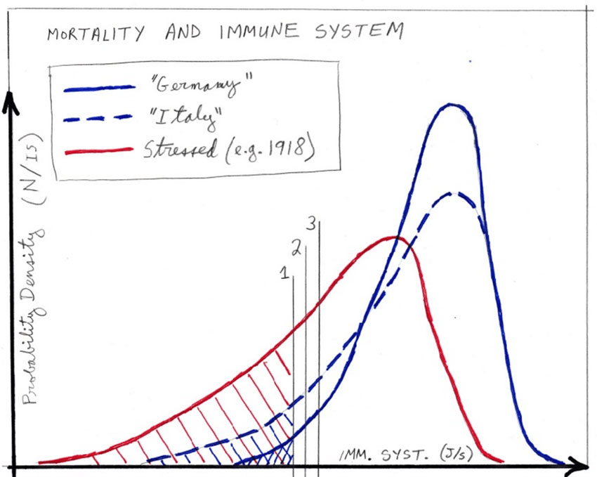
Figure 4 illustrates three hypothetical distributions of RS values,
in three different populations of equal size.
Here,
-
"Germany"
(solid-blue line) is for a current Western population, not having a
particularly large elderly population
-
"Italy" (dashed-blue line) is
for a current Western population having a large elderly population
-
"Stressed"
(solid-red line) is for a population of individuals
subjected to high metabolic (or health) stress, such as
might have been the case in 1918 England
Such health stress can arise from nutritional deficiency, essential
nutrient or vitamin efficiency, high levels of environmental
stressor-agents, toxins, or pathogens, shelter deficiency ("fuel
poverty"), oppressive working conditions, social-dominance
oppression, substance abuse causing organ damage, and so on.
There
is a vast literature on these factors.
As one anchor point, see: Sapolsky (2015); Sapolsky (2005).

Figure 4:
Probability densities of RS values, for three populations
of equal size but differing in health-stress levels
and health
vulnerabilities, as explained in the text.
The three vertical lines,
drawn in pencil and labeled
"1", "2" and "3", show three different
virus-specific
values of RSv, as explained in the text.
The hatched
areas are the fractions (of total area)
representing the mortality
fractions for the
less virulent virus having RSv value
labeled "1".
© D. G. Rancourt
In this model, therefore, comparative mortality between populations,
for a given viral pathogen, is determined by the different health
states (distributions of RS values of the individuals) of the
compared infected populations.
This is for the full cycle of infection and recovery.
It says little
about both the death rates on a daily basis and age distributions,
which depend on the natural or forced spread of the infection, which
in turn is not necessarily uniform in time and space but rather can
target particular segments of the population, such as people
confined in institutions.
Furthermore, the distribution of RS values for a given population
can change significantly during the course of an epidemic, if
vulnerable segments are subjected to additional health stressors,
for example.
All-cause mortality analysis of COVID-19
In light of the above background and conceptual tools, we can now
examine data for COVID-19, to date.
For good reason (as per above),
we ignore death-attributed data and model deconvolutions of P&I
deaths versus other deaths deemed to be seasonal for reasons
unrelated to the seasonal viral pathogens. We concentrate on
all-cause mortality, by week.
All-cause mortality is not susceptible to bias, and is currently
available for several jurisdictions.
We use the raw data without any
manipulation, and we do not modify the data to "correct" for changes
in total population, or for changes in age structure of a
population.
For the data, we rely on the CDC (USA), national institute data for
England and Wales, and the graphical compilations of the EuroMOMO
hub. We use only the latest weeks that are reported as complete
(">100%", CDC) or reported to be of sufficient quality to publish.
Unfortunately, some jurisdictions such as Canada can be
characterized as slow and refractory to requests.
Figure 5 shows all-cause mortality by week for England and Wales,
starting in 2010. The sudden single-week drops are book-keeping and
death-certification-delay inconsistencies, which are counted in the
following week(s).
The red vertical line indicates the date at which
the WHO declared the pandemic.
In declaring the pandemic, the WHO Director-General, Tedros Adhanom,
put it this way, among other things: 2
In the days and weeks ahead, we expect to see the number of cases,
the number of deaths, and the number of affected countries climb
even higher. [...]
And we have called every day for countries to take urgent and
aggressive action. We have rung the alarm bell loud and clear. [...]
This is not just a public health crisis, it is a crisis that will
touch every sector - so every sector and every individual must be
involved in the fight.
I have said from the beginning that countries must take a
whole-of-government, whole-of-society approach, built around a
comprehensive strategy to prevent infections, save lives and
minimize impact. [...]
I remind all countries that we are
calling on you to activate and scale up your emergency response
mechanisms; Communicate with your people about the risks and how
they can protect themselves - this is everybody's business;
Find, isolate, test and treat every case and trace every
contact; Ready your hospitals; [...]
Adhanom's words either were the most remarkable public health
forecast ever made for England and Wales (and many jurisdictions in
the world, see below), or something else might explain the sharp
peak in all-cause mortality that immediately followed his
declaration.

Figure 5:
All-cause mortality by week
for England and Wales,
starting in 2010.
The sudden single-week drops are book-keeping and
death-certification-delay inconsistencies, which are counted
in the
following week(s). The red vertical line indicates
the date at which
the WHO declared the COVID-19 pandemic.
© D. G. Rancourt
Importantly, the total number of winter-burden all-cause "excess"
deaths for the season ending in 2020 (area above the summer
baseline) is not statistically larger than for past years, and it
remains to be seen how low the summer 2020 trough will be.
What can be called "the COVID peak" is a narrow feature (Figure 5).
Relative to the summer baseline, the full-width at half-maximum of
the peak is approximately 5 weeks. It has the distinction of being
late in the infectious season, and of climbing far above the broader
winter-burden hump.
This "COVID peak" is a unique event in the epidemiological history
of England and Wales.
Does this unique feature arise from an
unusually novel viral pathogen, or does it arise from the unique,
unprecedented and massive government response to the WHO declaration
of a pandemic?
Note that such a "COVID peak" does not imply intrinsic virulence of
the virus. It only means that the deaths of vulnerable persons, or
persons made vulnerable, occurred in a short time span.
For example,
those who would have died in the next few or more weeks or months
can have their deaths accelerated by human intervention, or those
who are still recovering from a viral infection can be thrust into
more precarious and stressful living conditions.
An analogous "COVID peak" occurred in the EuroMOMO hub data for
Europe (Figure 6).
Here again, the total number of winter-burden
all-cause excess deaths for the season ending in 2020 (area above
the summer baseline) is not statistically larger than for past
years, and the date of declaration of the pandemic is shown by a
vertical red line.

Figure 6:
All-cause mortality by week
EuroMOMO hub data for Europe,
accessed on 1 June 2020.
The date of declaration of the pandemic
is
shown by a vertical red line.
© D. G. Rancourt
What looked like a concluding and "mild" 2020 season turned into a "COVID
peak" immediately after the WHO declared the pandemic.
Let us next move to the USA, where both national and state-by-state
current data is readily available, thanks to the CDC.
Figure 7 shows all-cause mortality by week for the USA, starting in
2014. Here the summer baseline is at approximately 46,000 to 52,000
deaths per week, increasing with the increase in total population.
The red vertical line indicates the date at which the WHO declared
the COVID-19 pandemic.

Figure 7:
All-cause mortality by week for the USA,
starting in 2014.
The red vertical line
indicates the date at which the WHO
declared the COVID-19 pandemic.
The hatched or gray-fill areas represent
the all-cause winter-burden
deaths
for each year.
© D.G. Rancourt
Here, again, we see that the total number of winter-burden all-cause
deaths for the season ending in 2020 (area above the summer
baseline) is not statistically larger than for past recent years.
There is no evidence, purely in terms of number of seasonal deaths,
to suggest any catastrophic event or exceptionally virulent
pathogen. There was no "plague". The winter burden, in these years,
is consistently in the range of approximately 6% to 9% of total
yearly all-cause mortality, and the year to year variations are
typical of historic variations.
On the other hand, there is again a "COVID peak", which has the
following unique features:
-
It is remarkably sharp or narrow, having a full-width at
half-maximum of the peak, relative to the summer baseline, of
approximately only 4 weeks. By comparison, the sharp peaks in the
infectious seasons ending in 2015 and 2018 have such full-widths of
14 and 9 weeks, respectively.
-
It occurs later in the infectious season than any other large sharp
peak ever seen for the USA, surging after week-11 of 2020.
-
It surge occurs immediately after the WHO declared the pandemic, in
perfect synchronicity, as seen in both Europe, and England and
Wales, which are an ocean apart from the USA.
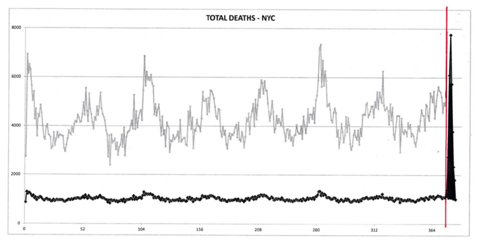
The "COVID peak" in the USA data arises from "hot spots", such as
New York City (NYC). Figure 8 shows the all-cause mortality by week
for NYC, starting in 2013.
The red vertical line indicates the date
at which the WHO declared the COVID-19 pandemic.

Figure 8:
All-cause mortality by week for NYC,
starting in 2013, in
black. The red vertical line
indicates the date at which the WHO
declared the COVID-19 pandemic.
The grey line is simply
the same data on a vertically expanded
and shifted scale, for
visualization.
© D.G. Rancourt
The NYC data makes no epidemiological sense whatsoever.
The "COVID
peak" here, on its face, cannot be interpreted as a normal viral
respiratory disease process in a susceptible population. Local
effects, such as importing patients from other jurisdictions or high
densities of institutionalized or housed vulnerable people, must be
in play, at least.
What is also striking is that some of the largest-population states
in the USA, having large numbers of measured and reported cases, and
large numbers of individuals with the antibodies, do not show a "COVID
peak". (Characteristic antibodies are produced and stored in the
bodies of individuals who were infected and recovered following
their immune responses. For example, see the antibody field study
for California done by Bendavid et al., 2020).
This is shown for California in Figure 9, and for Texas in Figure
10.

Figure 9:
All-cause mortality by week for California,
starting in
2013. The red vertical line
indicates the date at which the WHO
declared the COVID-19 pandemic.
The hatched or gray-fill areas represent
the all-cause winter-burden
deaths for each year.
© D.G. Rancourt

Figure 10:
All-cause mortality by week for Texas, starting in 2013.
The red vertical line indicates the date at which
the WHO declared
the COVID-19 pandemic.
The hatched or gray-fill areas represent the
all-cause winter-burden deaths for each year.
© D.G. Rancourt
Also, none of the seven states that did not impose a lockdown (Iowa,
Nebraska, North Dakota, South Dakota, Utah, Wyoming, and Arkansas)
have a "COVID peak".
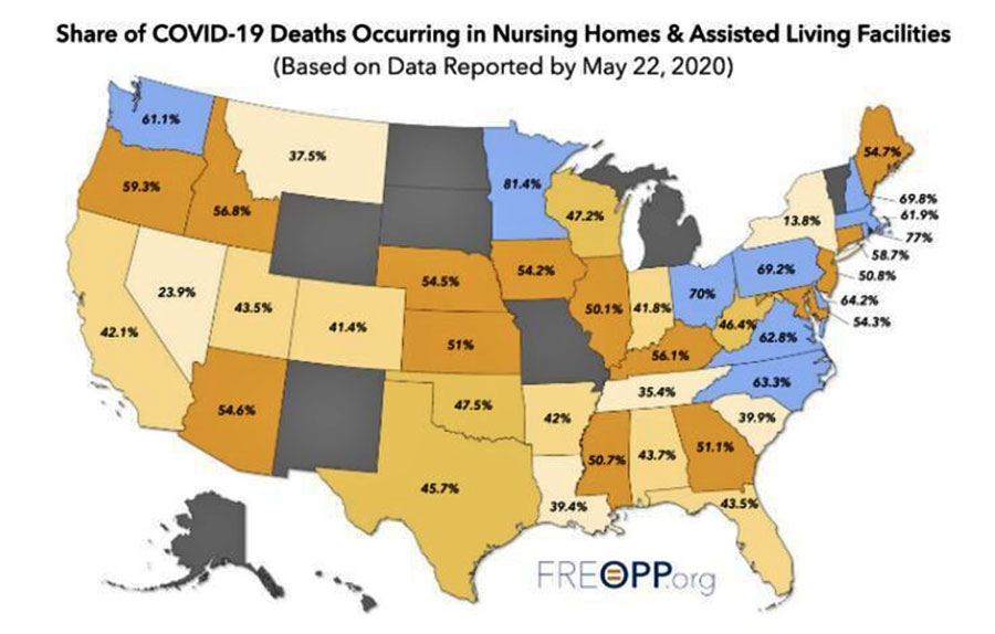
The presence of a "COVID peak" is positively correlated with the
share of COVID-19-assigned deaths occurring in nursing homes and
assisted living facilities, as per this map:

Interpreting the all-cause mortality "COVID peak"
Given the uniqueness of the all-cause mortality "COVID peak":
-
Its sharpness, with a full-width at half-maximum of only
approximately 4 weeks;
-
Its lateness in the infectious-season cycle, surging after week-11
of 2020, which is unprecedented for any large sharp-peak feature;
-
The synchronicity of the onset of its surge, across continents, and
immediately following the WHO declaration of the pandemic;
-
and its USA state-to-state absence or presence for the same viral
ecology on the same territory being correlated with nursing home
events and government actions rather than any known viral strain
discernment.
Given the above review of knowledge about seasonal viral respiratory
diseases:
-
The robustly persistent and regular winter-burden patterns of
all-cause mortality, across the modern era of epidemiology, and
across nations in two hemispheres;
-
The newfound (2010) understanding that transmissivity is controlled
by absolute humidity, and that the transmission vector is small
aerosol particles taken deeply into the lungs;
-
The increasing recognition of metabolic energy budgeting as the
paradigm for understanding death from infectious diseases with
comorbidity conditions, while recognizing that the immune system has
hierarchical control over metabolic energy budgeting, second only to
cognition of external imminent danger;
-
and the increasing understanding of the dominant role of metabolic
stress (including stress cognition, perceived stress) in depressing
immune system response capacity.
I postulate that the "COVID peak" represents an accelerated mass
homicide of immune-vulnerable individuals, and individuals made more
immune-vulnerable, by government and institutional actions, rather
than being an epidemiological signature of a novel virus,
irrespective of the degree to which the virus is novel from the
perspective of viral speciation.
Finally, my interpretation of the "COVID peak" as being a signature
of mass homicide by government response is supported by several
institutional documents, media reports, and scientific articles,
such as the following examples.
Two scientific articles are
on-point:
-
Hawryluck et al. (2004), on posttraumatic stress disorder (PTSD)
arising from medical quarantine.
-
Richardson et al. (2020), on statistical proof that mechanical
ventilators killed critical COVID-19 patients.
Media articles and institutional memos
"New study finds nearly all coronavirus patients put on ventilators
died", News Break | The Hill 04-23, 23 April 2020.
"New health care data suggests that almost half of all coronavirus
patients placed on ventilators die, first reported by CNN.
The data
was gathered at Northwell Health, New York state's largest hospital
system. It revealed that about 20 percent of COVID-19 patients
passed away, and 88 percent of those placed on ventilators died."
"Daughter blames 'chaos' of COVID-19 pandemic for mother's rapid
decline", by Arthur White-Crummey, Regina Leader-Post, 29 May 2020.
"Sue Nimegeers's mother never had COVID-19, but she still counts her
as a victim of the disease. 'She never tested positive, but the
chaos of the pandemic itself around us, we feel, took her
from us just way too soon,' Nimegeers told the board of the Saskatchewan
Health Authority (SHA) on Friday."
"'Deeply disturbing' report into Ontario care homes released", BBC,
27 May 2020.
"Mr Ford said a full investigation has been launched into the
allegations, which included claims that facilities smelt of rotten
food, infested with cockroaches and flies, and that elderly people
were left for hours 'crying for help with staff not responding'."
"Nothing can justify this destruction of people's lives", Yoram
Lass, former director of Israel's Health Ministry, on the hysteria
around Covid-19, sp!ked, 22 May 2020.
"Yoram Lass: It is the first epidemic in history which is
accompanied by another epidemic - the virus of the social networks.
These new media have brainwashed entire populations. What you get is
fear and anxiety, and an inability to look at real data. And
therefore you have all the ingredients for monstrous hysteria.
It is
what is known in science as positive feedback or a snowball effect.
The government is afraid of its constituents. Therefore, it
implements draconian measures.
The constituents look at the
draconian measures and become even more hysterical."
"Cuomo downplays calls for federal probe into nursing home
coronavirus deaths: 'Ask President Trump' ", by Andrew O'Reilly |
Fox News, 20 May 2020.
"New York Gov. Andrew Cuomo on Wednesday brushed off calls for the
Department of Justice to open an investigation into the massive
number of deaths in the state's nursing homes during the coronavirus
pandemic - claiming he was only following guidelines from the Trump
administration and Centers for Disease Control and Prevention.
While
no formal probe has been announced, the speculation comes amid
scrutiny of his March 25 directive that required nursing homes to
take on new patients infected with COVID-19."
DATE: March 25, 2020
TO: Nursing Home Administrators, Directors of Nursing, and Hospital
Discharge Planners
FROM: New York State Department of Health Advisory: Hospital
Discharges and Admissions to Nursing Homes (Removed from:
coronavirus.health.ny.gov)
"During this global health emergency, all NHs must comply with the
expedited receipt of residents returning from hospitals to NHs.
Residents are deemed appropriate for return to a NH upon a
determination by the hospital physician or designee that the
resident is medically stable for return. [...]
No resident shall be
denied re-admission or admission to the NH solely based on a
confirmed or suspected diagnosis of COVID-19.
NHs are prohibited
from requiring a hospitalized resident who is determined medically
stable to be tested for COVID-19 prior to admission or readmission."
"Nursing Homes & Assisted Living Facilities Account for 42% of
COVID-19 Deaths: A startling statistic has profound implications for
the way we've managed the coronavirus pandemic", by Gregg Girvan,
FREOPP, 7 May 2020.
"Based on a new analysis of state-by-state COVID-19 fatality
reports, it is clear that the most underappreciated aspect of the
novel coronavirus pandemic is its effect on a specific population of
Americans: those living in nursing homes and assisted living
facilities."
"Guilty - Of Breathing", by Tony Heller, Tony Heller YouTube
Channel, 24 May 2020.
"Lockdowns were sold months ago on the idea of 'flattening the
curve'. In most places there never was much of a curve to flatten,
yet the lockdowns are still in place.
Tens of millions are now
having their lives destroyed - for the crime of breathing."
"The 'massacre' of Italy's elderly nursing home residents: Covid-19
patients in Italy's virus epicentre of Lombardy were transferred to
nursing homes by an official resolution with catastrophic
consequences", by Maria Tavernini and Alessandro Di Rienzo, TRT
World, 20 April 2020.
"Hosting Covid-19 patients in nursing homes was like lighting a
match in a haystack."
"Coronavirus Update: How shoring up hospitals for COVID-19
contributed to Canada's long-term care crisis", by Jessie Willms and
Hailey Montgomery, Globe & Mail, 20 May 2020.
"Most of the nursing- and retirement-home residents who have
succumbed to COVID-19 in Canada died inside the virus-stricken,
understaffed facilities as hospital beds sat empty."
"There Is No Evidence Lockdowns Saved Lives. It Is Indisputable They
Caused Great Harm", by Briggs, wmbriggs.com, 14 May 2020.
"In the end, it does not come down to country- or even city-level
statistics. It comes down to people. Each individual catches the bug
or not, lives or dies.
Not because of their country, but because of
themselves, their health, their circumstances.
Any given individual
might have benefited from self-quarantine and loss of job. Just as
any given individual might have come to a bad end from a lockdown."
"Hospitals get paid more to list patients as COVID-19", by Tom
Kertscher, POLITIFACT, 21 April 2020.
"It's standard for Medicare to pay a hospital roughly three times as
much for a patient who goes on a ventilator, as for one who doesn't.
Medicare is paying a 20% add-on to its regular hospital payments for
the treatment of COVID-19 victims. That's a result of a federal
stimulus law."
"CDC: 80,000 people died of flu last winter in U.S., highest death
toll in 40 years", by Associated Press, STAT News, 26 September
2018.
"An estimated 80,000 Americans died of flu and its complications
last winter - the disease's highest death toll in at least four
decades.
The director of the Centers for Disease Control and
Prevention, Dr. Robert Redfield, revealed the total in an interview
Tuesday night with The Associated Press."
Footnotes
-
'The immune system:
Cells, tissues, function, and disease', medically reviewed by
Daniel Murrell, MD on January 11, 2018 - Written by Tim Newman,
at
medicalnewstoday.com, accessed on 1 June, 2020.
-
WHO Director-General's opening remarks at the media briefing on
COVID-19 -
11 March 2020
Scientific
references
-
Alimpiev,
Egor (2019) "Rethinking the Virus Species Concept", dated 15
March 2019, posted to
stanford.edu
-
Baccam, P.
et al. (2006) "Kinetics of Influenza A Virus Infection in
Humans",
Journal of Virology Jul 2006, 80 (15) 7590-7599;
DOI: 10.1128/JVI.01623-05
-
Bajgar et
al. (2015) "Extracellular Adenosine Mediates a Systemic
Metabolic Switch during Immune Response",
PLoS Biol 13(4): e1002135.
-
Bendavid et
al. (2020) "COVID-19 Antibody Seroprevalence in Santa Clara
County, California",
medRxiv 2020.04.14.20062463
-
Brooke, C.
B. et al. (2013) "Most Influenza A Virions Fail To Express
at Least One Essential Viral Protein",
Journal of Virology Feb 2013, 87 (6) 3155-3162; DOI:
10.1128/JVI.02284-12
-
Dowell, S.
F. (2001) "Seasonal variation in host susceptibility and
cycles of certain infectious diseases",
Emerg Infect Dis. 2001;7(3):369-374.
doi:10.3201/eid0703.010301
-
Haas, C.N.
et al. (1993) "Risk Assessment of Virus in Drinking Water",
Risk Analysis, 13: 545-552.
doi:10.1111/j.1539-6924.1993.tb00013.x
-
Harper, G
J. (1961) "Airborne micro-organisms: survival tests with
four viruses",
The Journal of Hygiene,
vol. 59,4: 479-86. doi:10.1017/s0022172400039176
-
Hawryluck,
L. et al. (2004) "SARS control and psychological effects of
quarantine, Toronto, Canada",
Emerging Infect Dis., vol. 10,7: 1206-12.
doi:10.3201/eid1007.030703
-
HealthKnowlege-UK (2020) "Charter 1a - Epidemiology:
Epidemic theory (effective & basic reproduction numbers,
epidemic thresholds) & techniques for analysis of infectious
disease data (construction & use of epidemic curves,
generation numbers, exceptional reporting & identification
of significant clusters)",
HealthKnowledge.org.uk, accessed on 2020-04-10.
-
Hsieh, Y.C.
et al. (2006) "Influenza pandemics: past, present and
future",
J Formos Med Assoc. 105(1):1-6.
doi:10.1016/S0929-6646(09)60102-9
-
Langmuir,
A.D. (1976) "William Farr: Founder of Modern Concepts of
Surveillance",
International Journal of Epidemiology, Volume 5,
Issue 1, March 1976, Pages 13-18,
-
Locey and
Lennon (2016) "Scaling laws predict global microbial
diversity",
Proceedings of the National Academy of Sciences,
May 2016, 113 (21) 5970-5975; DOI: 10.1073/pnas.1521291113
-
Lowen, A.
C. et al. (2007) "Influenza Virus Transmission Is Dependent
on Relative Humidity and Temperature",
PLoS Pathog 3(10): e151.
-
Lui, K.J.,
Kendal, A.P. (1987) "Impact of influenza epidemics on
mortality in the United States from October 1972 to May
1985",
Am J Public Health,
77(6):712-716. doi:10.2105/ajph.77.6.712
-
Marti-Soler, H. et al. (2014) "Seasonal Variation of Overall
and Cardiovascular Mortality: A Study in 19 Countries from
Different Geographic Locations",
PLoS ONE, 9(11): e113500.
-
Rancourt,
D.G. (2020), "Masks Don't Work: A review of science relevant
to COVID-19 social policy", Technical Report,
Research Gate, 10 April 2020, DOI:
10.13140/RG.2.2.14320.40967/1
-
Richardson,
S. et al. (2020) "Presenting Characteristics, Comorbidities,
and Outcomes Among 5700 Patients Hospitalized With COVID-19
in the New York City Area",
JAMA.
323(20):2052-2059. doi:10.1001/jama.2020.6775
-
Sapolsky
(2005) "The Influence of Social Hierarchy on Primate
Health",
Science, 29 April 2005, vol. 308, pages 648-652.
DOI: 10.1126/science.1106477
-
Sapolsky
(2015), "Stress and the brain: individual variability and
the inverted-U",
Nature Neuroscience,
October 2015, vol. 18, no. 10, pages 1344-1346. doi:
10.1038/nn.4109.
-
Shaman, J.
et al. (2010) "Absolute Humidity and the Seasonal Onset of
Influenza in the Continental United States", PLoS Biol 8(2):
e1000316.
https://doi.org/10.1371/journal.pbio.1000316
-
Simonsen,
L. et al. (1997) "The impact of influenza epidemics on
mortality: introducing a severity index",
Am J Public Health. 87(12):1944-1950.
doi:10.2105/ajph.87.12.1944
-
Straub RH.
(2017) "The brain and immune system prompt energy shortage
in chronic inflammation and ageing",
Nat Rev Rheumatol.
13(12):743-751. doi:10.1038/nrrheum.2017.172
-
Viboud, C.
et al. (2010) "Preliminary Estimates of Mortality and Years
of Life Lost Associated with the 2009 A/H1N1 Pandemic in the
US and Comparison with Past Influenza Seasons",
PLoS currents, vol.
2 RRN1153. 20 Mar. 2010, doi:10.1371/currents.rrn1153
-
Viboud C.
et al. (2006) "Transmissibility and mortality impact of
epidemic and pandemic influenza, with emphasis on the
unusually deadly 1951 epidemic",
Vaccine. 24(44-46):6701-6707.
doi:10.1016/j.vaccine.2006.05.067
handelgroup.publichealth.uga.edu
-
Viboud, C.
et al. (2005) "Multinational Impact of the 1968 Hong Kong
Influenza Pandemic: Evidence for a Smoldering Pandemic",
The Journal of Infectious
Diseases, Volume 192, Issue 2, 15 July 2005, Pages
233-248,
-
Yang, W. et
al. (2011) "Concentrations and size distributions of
airborne influenza A viruses measured indoors at a health
centre, a day-care centre and on aeroplanes",
Journal of the Royal Society,
Interface. 2011 Aug;8(61):1176-1184. DOI:
10.1098/rsif.2010.0686.
-
Yezli, S.,
Otter, J.A. (2011) "Minimum Infective Dose of the Major
Human Respiratory and Enteric Viruses Transmitted Through
Food and the Environment",
Food Environ Virol
3, 1-30.
-
Zwart, M.
P. et al. (2009) "An experimental test of the independent
action hypothesis in virus-insect pathosystems",
Proc. R. Soc. B.
2762233-2242
|